Fibrous Dysplasia- a case report
Dr Biplob Kumar Halder, Prof. Dr. Shahidul Islam, Dr. Jan Mohammad, Dr. Umme Iffat Siddiqua, Dr. Sohel Abdullah
A 43years old gentleman, an executive of an Airline Office came to United Hospital orthopedic department with history of pain in left hip joint. Patient was normotensive and non-diabetic. He was then sent to the Radiology & Imaging Department for MRI of pelvis. On STIR images subtle hyperintensity was noted in left iliac bone and along the left acetabular margin. Then he was asked to do a CT scan of pelvis which showed expansile irregular lytic and sclerotic lesion in left iliac bone and along the left acetabular margin. However the joint space and adjacent soft tissues were unremarkable. Then our provisional diagnosis was fibrous dysplasia and the patient was then further advised to do a confirmatory bone biopsy (CT guided) which confirmed Fibrous Dysplasia.
| |
|
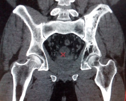
CT Image: Fibrous Dysplasia of left hip bone
|
|
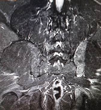
MRI Image: Fibrous Dysplasia of left hip bone
Fibrous dysplasia is a non-neoplastic disorder where normal bone marrow is replaced with fibrous tissue resulting in formation of bone that is weak and prone to expansion. It can affect any bone in the body but most often occurs in the:
The significant complications are fracture, deformity, functional impairment and pain. This disease can affect single bone (monostotic) or multiple bones (polyostotic) and may occur in isolation or in a complex genetic disorder termed McCune-Albright Syndrome. More rarely fibrous dysplasia may be associated with intramuscular myxoma in a condition termed Mazabraud's Syndrome.