Removal of any abnormal bony growth around the large joint or long bone is urgent- A Case report
Dr.A.H.M Rezaul Haque, Dr. Masum Billah
Aneurysm /pseudo-aneurysm or nerve compression may developed at anywhere in the course of neurovascular supply (mostly along the long bone or around large joints) due to injury of the vascular wall by any abnormal bony growth or exostosis.It is rational to do operation after closure of epiphysis. But in popliteal space it should be done as early as possible, waiting for closure of epiphysis may create new complications (Transformation of carcinoma or cosmetic purpose). So, early detection and management can be beneficial for the patients.
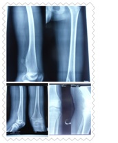
Case history: A 14 years 3 months old young boy was admitted in UHL through OPD-1 as a known case of osteochondroma/bony growthon lower 1/3rd of left femur for 7 months. Suddenly he developed flexion deformity, pain, swelling on popliteal fossa and unable to move left knee joint for 8 days. On X-ray left knee joint revealed spiky bony projection/growth in postero-medial aspect and lower 1/3rd of left femur. He is also having H/O bronchial asthma.
On local examination of left lower limb, there was a swelling in postero-medial aspect and lower 1/3rd of left thigh which is hard, fixed and tender. Redness was present. Local temperature was raised. ROM of left knee joint was painful and restricted. Left ADP was palpable. He can move toes and ankle actively. No neurological deficit was found.MRI of left knee: Known case of osteochondroma with suspected malignant transformation/inflammatory

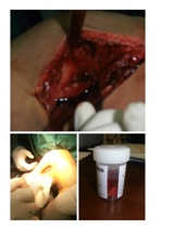
 After proper counseling with patient’s attendant about the swelling and its treatment outcome, patient underwent Excisional biopsy (osteochondroma??) of left knee under G/A on 30/11/2015 and tissue sent for histopathology.
Findings of Operation:There was a huge sac surrounded by a thin membrane with full of clotted blood (Half of the clot was removed) extended from lower 1/3rd of left femur to back of popliteal fossa at medial site. A bony stalk (aprrox 3cm X 2cm) seen above the left distal femoral condyle, which was excised and sent for histopathological examination.His post-operative period was uneventful.
He was in regular follow up, swelling was reduced, can move the left knee actively, distal circulation of left lower limb was normal with neurological impairment. After four months DopplerUSG of left popliteal fossa done and revealed huge pulsatile mass was present and advised to consult with vascular surgeon.